Key Clinical Message
Eschar formation is a potential sequela of burn injuries. Definitive management may include escharectomy and eschar debridement. After eschar removal, the wound can be covered with a skin graft or reepithelialization. For prolonged refractory eschar on the fingertips, topical use of rb-bFGF after debridement can achieve an optimal outcome.
Keywords
Accumulative eschar, fibroblast growth factor, granulation wound, hand burn.
Introduction
Eschar is composed of dead tissue and dried secretions from a skin wound following a burn or an infectious disease on the skin. The eschar provides temporary coverage of and protection to the wound. An eschar normally persists for less than a month before sloughing off or dissolving itself [1]. However, we encountered a patient with prolonged refractory accumulative eschar formation on her fingertips and finger pulps that remained for more than 2 years after a burn injury.
Case Report
A 28-year-old female presented with eschar crust on her fingertips and finger pulps. The patient had experienced a burn injury on the hand from an alcohol flame 2 years prior. The majority of the wounds healed well after topical ointment treatment. However, there were several areas of the fingertips and finger pulps where the new skin was thin, red, fragile, and sensitive. Six months after the injury, the thin skin in these areas was replaced by excretive granulation tissues. Initially, the secretions dried and a thin layer of crust formed. However, the crust thickened and led to formation of proliferating eschars.
The patient experienced mild discomfort but did not have pain in the affected areas. The accumulated thick eschar affected the function of the patient’s hands and interfered with everyday activities. To remove the eschars, the patient soaked her hand in water and removed the crust manually after the eschars had softened. The process was repeated every 1 to 2 months, after the eschars reformed. The patient then removed the eschars again using the same method.
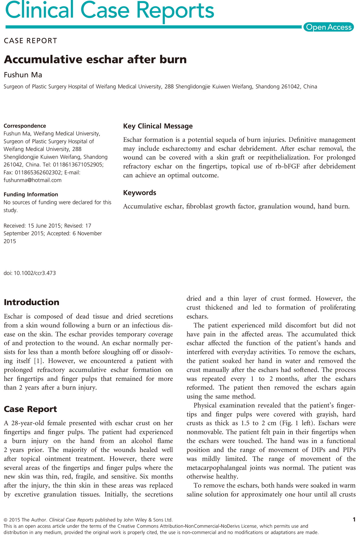
Physical examination revealed that the patient’s fingertips and finger pulps were covered with grayish, hard crusts as thick as 1.5 to 2 cm (Fig. 1 left). Eschars were nonmovable. The patient felt pain in their fingertips when the eschars were touched. The hand was in a functional position and the range of movement of DIPs and PIPs was mildly limited. The range of movement of the metacarpophalangeal joints was normal. The patient was otherwise healthy.
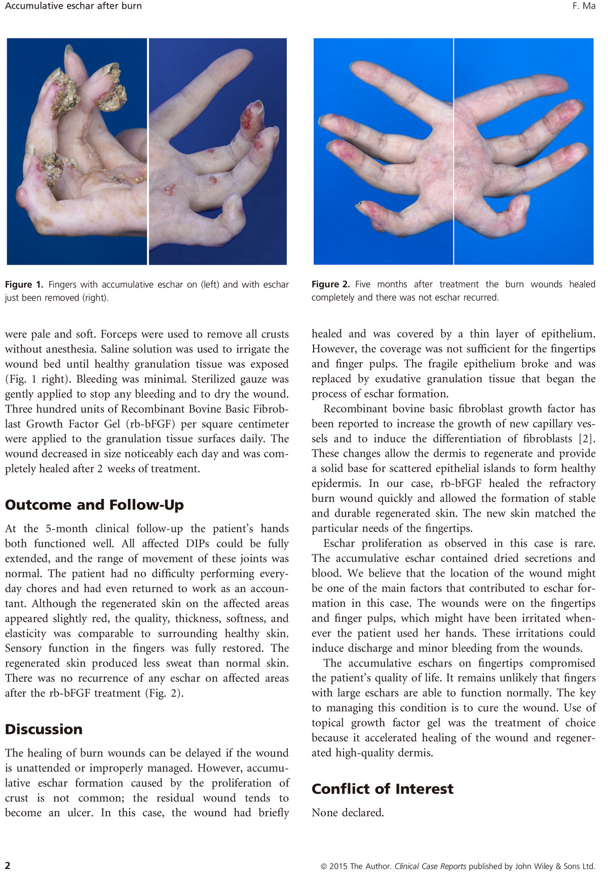
To remove the eschars, both hands were soaked in warm saline solution for approximately one hour until all crusts were pale and soft. Forceps were used to remove all crusts without anesthesia. Saline solution was used to irrigate the wound bed until healthy granulation tissue was exposed (Fig. 1 right). Bleeding was minimal. Sterilized gauze was gently applied to stop any bleeding and to dry the wound. Three hundred units of Recombinant Bovine Basic Fibroblast Growth Factor Gel (rb-bFGF) per square centimeter were applied to the granulation tissue surfaces daily. The wound decreased in size noticeably each day and was completely healed after 2 weeks of treatment………
以上是本文的缩减版本。全文已经于2015年12月9日发表在由美国Wiley出版社出自版的《临床病例报告杂志》(Clinical Case Report)。
下面是从杂志网站上下载的本文PDF版


